Article - What are the benefits of Tetris?
The theory is that Tetris thickens the cerebral cortex - part of the brain that plays a key role in memory, attention, perceptual awareness, thought,...


The cerebral cortex develops initially from the anterior neural plate, a specialised part of the embryonic ectoderm. As the neural plate folds to form the neural tube the cavity inside the tube develops the ventricular system. The neurons and glia of the nervous system then develop from the epithelial cells of the cavity wall.
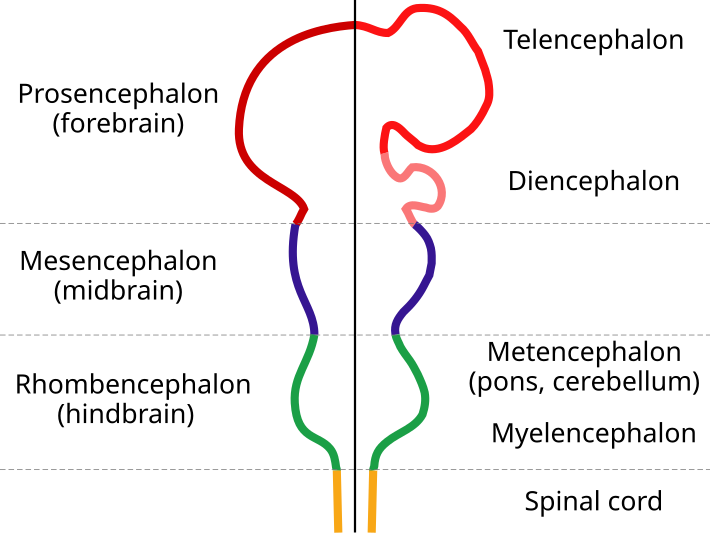
Embryonic Brain. Image courtesy of Wikimedia Commons. This image is in public domain.
This frontal part of the neural tube, known as the telencephalon (see above), is what gives rise to the cerebral hemispheres and cortex.
The development of the cerebral cortex (corticogenesis) involves four major stages:
Neurons of the cortex develop from the ventricular zone, adjacent to the ventricles, which at first contain "progenitor" cells (also known as germinal cells). These cells divide to produce glial and neuronal cells.
The glial fibres, which are created in the first division of progenitor cells, are oriented radially, projecting through the entire thickness of the cortex, from the ventricular zone to the outer, pial surface. These fibres provide a scaffold for the migration of neurons outwards from the ventricular zone.

Corticogenesis in a Wild-Type Mouse. Image courtesy of Wikimedia Commons. This image is in public domain.
The above image shows corticogenesis in a wild-type mouse. By using the radial glia fibres, differentiated progenitor cells migrate outwards (upwards in the image) to form the different layers of the cortex. The subplate neurons are the first to take thier place (yellow), followed by the cortical plate neurons (white to black), which migrate past the subplate.
The proteins laminin and reelin assist with this migration.
—————
The neurons of the cerebral cortex constitute the highest level of control in the hierarchy of the nervous system.